


https://www.researchgate.net/publication/378090792_Risk_of_Fall_Cognition_and_Static_Posture_in_Aging
I came across this very interesting article while listening to one of my chiropractic research podcasts. It reminded me of a conversation I had with a patient and her adult daughter who drove her to her appointment last year. The mother, in her early 80s, had moved closer to her daughter’s family and was trying to reestablish chiropractic care on a more consistent basis. She was a lifelong chiropractic patient, for episodic neck injuries and later on decided to stay on a preventive checkup schedule because she felt overall better and more balanced when she did so. During the history, her daughter chimed in on her mother’s comment that “ she has a tendency to be more clumsy and trip “ when she has not had a chiropractic check and adjustment in a while. The daughter was unfamiliar with chiropractic and simply curious about the correlation between the two. At the time, I share my 30 years of clinical experience in observing that correlation and the biological mechanisms connecting the chiropractic treatment on the neurological adjustment of proprioception and reflex time. I wish I had had that piece of research to add to the conversation.
The research led by a lesser known Brazilian colleague showed a correlation between two key measures of sagittal posture and the risk of fall: the degree of anterior neck shifting in relationship to the trunk, and the anterior angle of the ankle. Both indicating that the body is off its center of gravity and thus has less time to respond to stay upright before hitting the ground. Both posture indicators are exam findings that we observe and report, and both are factors that we aim to improve/correct with manual therapy and guidance on home activities and corrective exercises. One little golden nugget for me to take away from this research is that I can use it in conversation with patients when they question why I work on their lower extremities, especially their ankles and feet, when their primary problem seems to lie elsewhere. I almost often look at the lower extremity alignment when doing my initial evaluation and I will adjust them if needed, something that makes some folks a little testy when I ask them to take off their shoes and poke at one more body area. Now I can remind them that when I take on that additional endeavor at 5 pm, when their footsies are a little more “ aromatic”, it is indeed solely for their own benefit and backed by solid scientific research


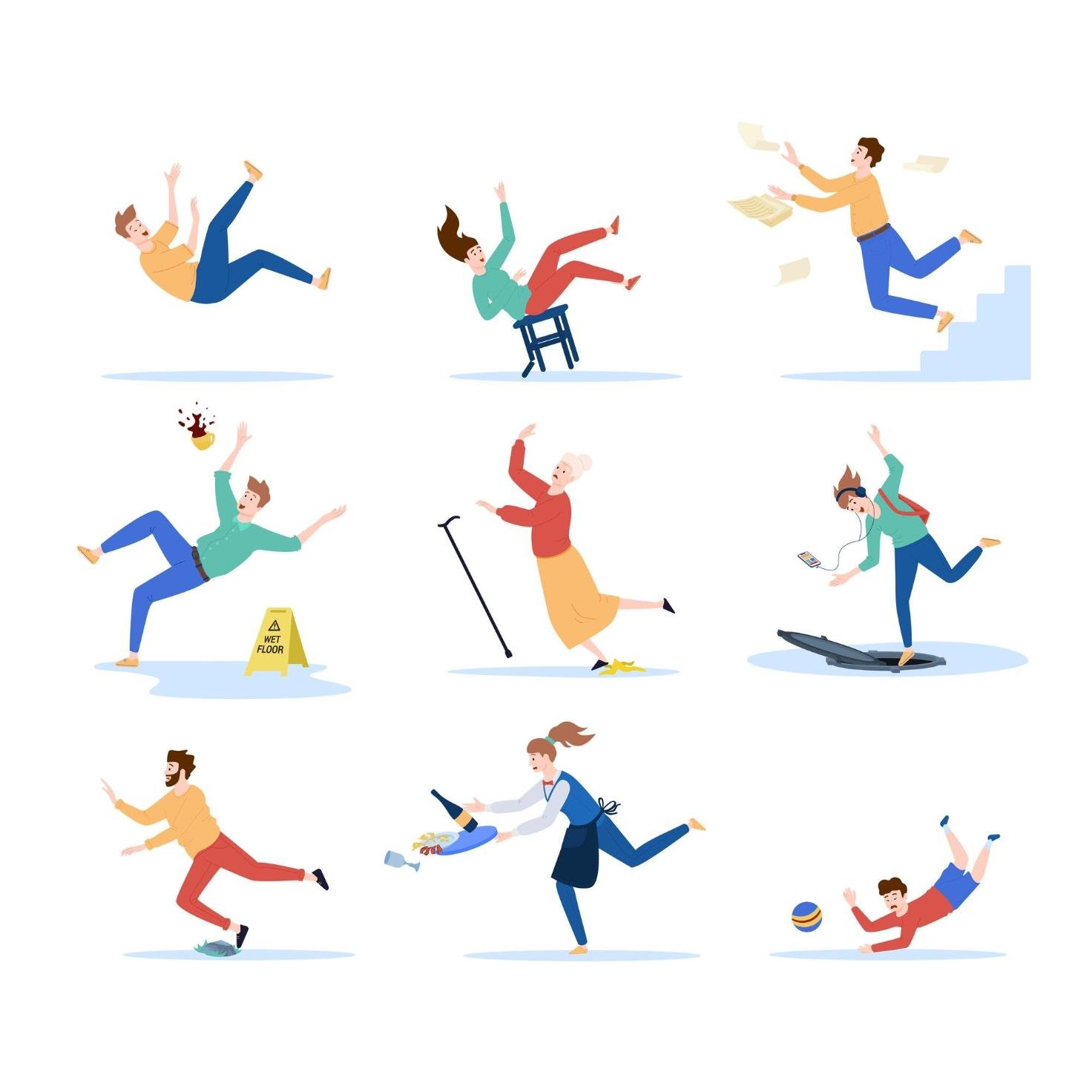
(photo courtesy Freepik)