Shoulder strength and stability: hug the ball

https://www.youtube.com/watch?v=YfpNnMjEsqw

When working with patients with shoulder girdle weakness and instability, whether from an acute injury or more of a chronic postural strain, there are a couple of foundational exercises that we will use as a starting point for strength and stability recovery. Those exercises can be done safely as a starting point with little to no risk of injury when performed correctly, and can be used as a progression tool to more aggressive strength building.

The "hug the ball" (or hug the pillows) exercise allows patients to recruit the scapula stabilizing muscles and prevent some of the chronic rounded shoulder and shoulder blade winging. The ball or the pillows allow for a counter resistance that can be light or intense, and allows for a slow eccentric release. The main technique to pay attention to is making sure to engage the deep interior blade muscles to squeeze the ball and not the biceps.

Shoulder strength and stability exercise: crocodile pushup

https://www.youtube.com/watch?v=frjhTszm3Cc&t=21s

On the same topic of shoulder strength and stability foundational exercises, this form of modified push-up is a great tool to be used as a stepping block when patients are extremely weak following a shoulder injury and not quite ready to do more traditional plank type exercises. Like the hug the ball exercise, it can be gradually made more challenging as the patient progresses in their recovery.

The "crocodile push-up" ensures that the elbows are facing the knees, keeping the elbows from flailing out, and the glenohumeral joint firmly centered in the socket. The patient will start doing the exercise from the knees up, and can bend as little as needed in order for the isometric hold to be tolerated for about 5 seconds before a slow release. The patient can eventually progressed to lower push up, longer hold time, and full plank starting position.

CHIROPRACTIC FOR PELVIC OUTLET CONSTIPATION: WHEN YOUR GUT AND YOUR BUTT DON’T LINE UP

CHIROPRACTIC FOR PELVIC FLOOR OUTLET CONSTIPATION: WHEN YOUR GUT AND YOUR BUTT DON’T LINE UP

After 30 years of practice I am glad to report that I learn new things almost every day and for certain every week. And recently I had 2 cases, one pediatric and one adult, that made me realize that certain subtypes of constipation are closely linked to distortion patterns in the spine, pelvis, and pelvic floor.

On my wish list is the ability to take the full pelvic floor clinical management review, which may happen in the next year when I'm able to make time and travel to the teaching outfiit I have selected. In the meantime, I have been hitting the books and doing short online webinars to try to get a better understanding of the pelvic floor anatomy, which in our profession and historical training has been a little bit of "no man's zone" of management, although I've come to appreciate over the last few years that it could often be the missing link between the trunk and the lower extremity.

The tongue-in-cheek title of this blog actually comes from a patient trying to describe her problem, as she had developed some unusual constipation, which really was the inability to release stools from the very lowest part of her rectum, following a pretty severe lumbar and gluteal injury. She felt foolish describing to me the sensation that her lower rectum did not align with her anus, with the sensation that it was almost sitting too far back onto the sacrum. But in reality she was describing exactly what was happening to her. It is a known clinical presentation that has been assigned an ICD 10 code (the official medical nomenclature of every medical condition for the sake of insurance coding and research): pelvic outlet constipation,K95.02.

Constipation is a term that covers a variety of symptoms that have an equal variety of causes. Pelvic outlet constipation is a very specific form of constipation that does not involve other traditional causes (low intestinal wall muscular activity, low fiber/bulk, internal stool dehydration). It is the inability of the lower rectum to empty out the stool content through the anal sphincter. Upwards digestion and motility and bulk are okay, but things are literally mechanically stuck trying to get out at the very end. Patients will describe a different sensation from other forms of constipation, sensing that the stool is very low, causing a lot of pressure in the rectum, and an urgent need to evacuate, but no matter how much they push they feel that things are stuck and not able to get out. This can cause the patient to spend an inordinate amount of time trying to push very hard, to the point of causing local injury in the form of hemorrhoids and occasional prolapse.

To make sense of this phenomenon you need to understand the basic anatomy of the lower colon in relationship to the pelvic floor and the pelvis itself.

The distal colon descends from the left lower abdomen into the front of the sacrum, where it's posterior to the anal opening. The muscular sphincter is surrounded by a complex set of pelvic floor muscles, which can move the sphincter further forward under voluntary control, thus holding stool in the rectum rather than letting them evacuate.

In order to let stool pass through, the pelvic floor muscles need to relax so that the sphincter can move a little further backwards and align itself with the rectum. In addition, the sphincter must be central in the pelvic floor, not shifted or pulled to one side, in order to have the easiest and smoothest stool evacuation.

If you look at the anatomical drawings of the pelvic floor muscle and the lower colon, you will notice that the pelvic floor muscles attach to the entirety of the bony pelvis, from the posterior part ( ilium and ischium, sacrotuberous ligament, tailbone) to the anterior part behind the pubic bone. In addition, all the pelvic floor muscles are innervated by by the lower lumbar segment and sacral segments.

The intimate neurological and structural connection of the pelvic floor to the lumbar spine and pelvis means that distortion patterns, trauma, repetitive strain of the lumbar spine and pelvis can result in some significant distortion of the muscular activity and tone of the pelvic floor, with the risk of chronically altering the central positioning of the sphincter in relationship to the lower rectum. This means that at the time of voluntary evacuation, it would take more effort and straining to bypass this suboptimal relationship between the lower large intestine and the outlet.

It occurred to me at the time I started writing this blog that I've probably been incidentally treating a lot of mild pelvic outlet constipation cases for a while, as a byproduct of my chiropractic treatments to the lumbar spine and pelvis. However it's only more recently that I've started paying attention more closely and in more detail to the structures of the posterior pelvic floor, which abuts structures we routinely palpate in the lower sacrum, inferior external rotators of the hip and sacral ligaments. I've also started asking the patients a few more pointed question about symptoms, which they may not volunteer, partially because these body parts are pretty private and partially because people don't make the connection between their G.I. symptoms and their lumbar pelvic symptoms.

To be effective, a chiropractic treatment in patients suffering from pelvic outlet constipation needs to incorporate several elements: addressing the structural alignment issues in the lumbar spine and especially in the pelvis; addressing the muscular imbalance of the external rotators of the hip, especially the most inferior group; addressing abnormal tone texture and scar tissue in the posterior pelvic ligaments (sacrotuberous and sacral spinous); and being able to give the patient basic information about how to do a proper pelvic floor stretch and strengthen based on which side is involved.

I'm also very fortunate to have access to several excellent pelvic floor specialty providers (physical therapist and occupational therapist) in my area of practice, who can take care of more direct pelvic floor manual therapy in patients who need it


K59.02

Outlet dysfunction constipationK59. 02 is a billable/specific ICD-10-CM code that can be used to indicate a diagnosis for reimbursement purposes. The 2024 edition of ICD-10-CM K59.


https://www.icd10data.com/ICD10CM/Codes/K00-K95/K55-K64/K59-/K59.02#:~:text=Outlet%20dysfunction%20constipation,-2016%202017%202018&text=Billable%2FSpecific%20Code-,K59.,ICD%2D10%2DCM%20K59.

Femoral neuritis: the "other sciatica"

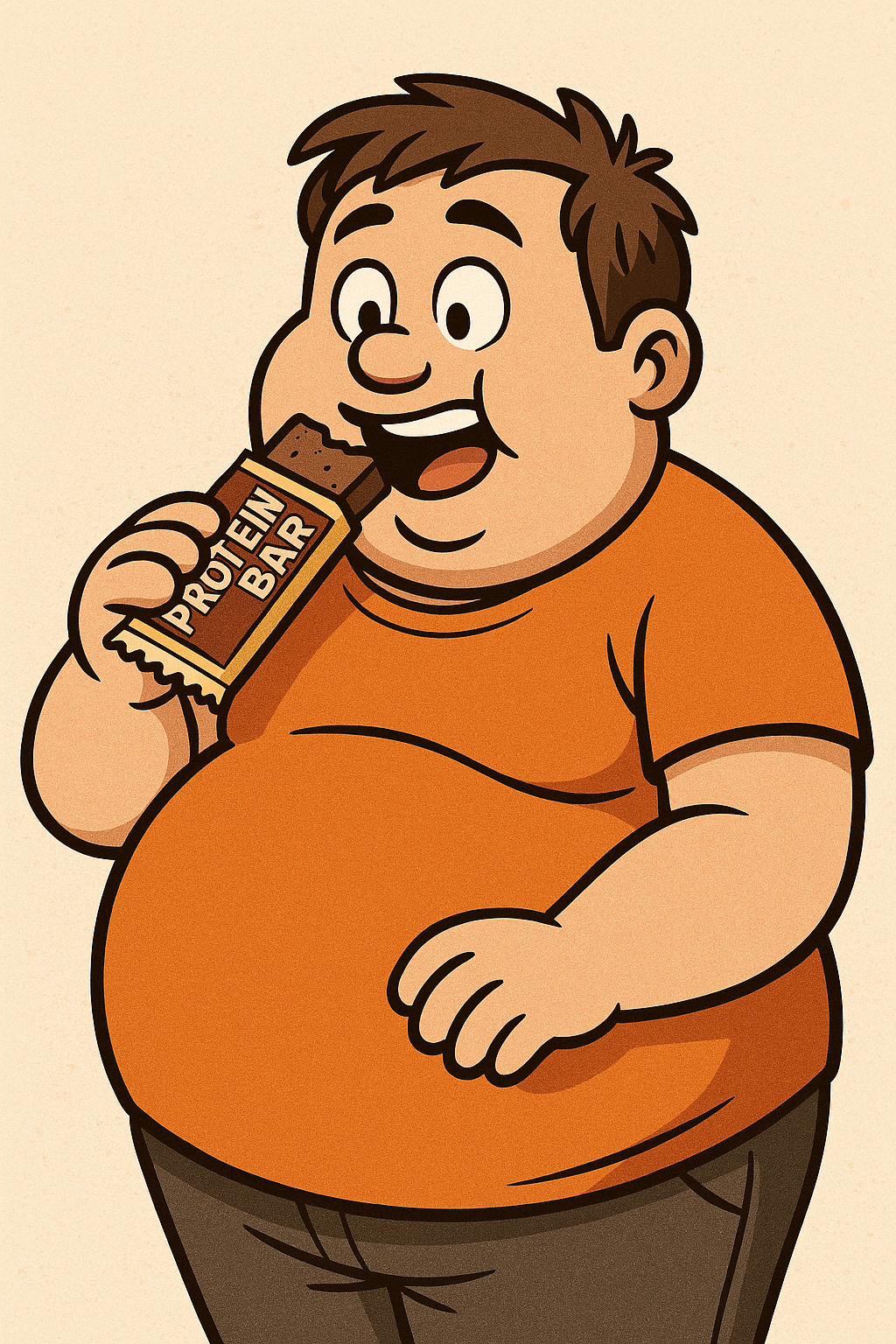
Femoral Neuritis

Sciatica enjoys quite a bit of popularity, and rightfully so. It is not technically a medical diagnosis but the description of symptoms encompassing pain in the leg, generally assumed to stem from the low back. The sciatic nerve originates at 5 levels spanning the last two lumbar vertebrae/discs, and the upper three sacral segments. It travels through several soft tissue structures in the buttock, down the posterior thigh, before splitting into two different branches at the knee, covering the lateral and posterior calf and foot. As such, sciatica describes referred pain affecting this distribution pattern.

Patients often use the term “sciatica” pretty liberally, to describe any sort of pain in the leg, including pain in the front of the thigh, which is not a sciatic nerve distribution. Enter its lesser known cousin, the femoral nerve.

Femoral neuritis is actually surprisingly common but getting little recognition ( it is still less common than sciatic neuritis). The femoral nerve originates in the mid lumbar spine and is made up from nerve roots from lumbar segments L2 through L4.  It travels in the front of the lumbar spine, deep in the abdomen, through the intersection of the two branches of the iliopsoas hip flexor muscles, through the groin and into the anterior and medial aspect of the thigh. It does not extend very far below the knee, unlike the sciatic nerve, which extends all the way into the foot.

The femoral nerve can be compressed in the lumbar spine, by a mid lumbar disc herniation, and just as commonly by myofascial injuries in the hip flexors. The patient will often present with unexplained groin, hip, thigh and medial knee pain and tingling. Unlike sciatic neuritis, many patients will not initially recognize  femoral neuritis as referred pain from the lumbar spine since it manifests in the front of the trunk and leg.

Femoral neuritis will be treated in the same manner as sciatic neuritis, based on the source / cause of the problem: chiropractic adjustments, myofascial release, corrective exercise, supportive therapies etc.

Caregiver injuries and transfer belts

https://www.youtube.com/watch?v=5GC_OETvnRc

This video is geared for a wide audience, not just for the professionals in the personal care industry. Many of us are finding ourselves in a season of life involving caring for loved ones at home with progressive leg weakness and balance issues. I have treated enough injuries sustained in that context to spur this brief video on how you can prevent many injuries using this simple tool and technique. If you are not sure on how to use a transfer belt, please bring your own to your appointment and I will be glad to give you a short tutorial

How to use orthotics in sandals?

It's a seasonal subject I find myself discussing a lot, as our most dedicated orthotics users are finding it cumbersome to keep their toes covered in sweltering conditions this year.

Orthotics can definitely be worn in sandals as long as you find the right sandal: it needs to have a deep enough removable footbed, and in most instances for patients with pronation, and adequate support along the medial ankle.

I keep a running list of the most common brands and models, which I'll happily email to patient's who requested. Since women's fashion is subject to an notoriously high turnaround, I do not post on the website for fear I would forget to update it periodically.

In the meantime, you can see in the video how easily and orthotic can be fitted in a good sandal.

https://www.youtube.com/watch?v=IjK0GV62QHM&t=5s

The 3 cats and three cows of the morning: how to get out of bed when your low back is really stiff and painful

We have had a lot of patients inquire about the best method to get out of bed and loosen up when they wake up with a lot of intense low back stiffness and pain. This is not an uncommon finding with patients who have underlying lumbosacral inflammation from new injuries or degenerative disc disease.

The brief video goes through 3 versions of the cat and cow, starting right on your back before getting out of bed, and gradually progressing through the seated version and into the traditional tabletop version. By the time you finally stand up, the pain is often 50% reduced. This allows patients, who normally dread getting out of bed because of the half hour of intense pain and stiffness, to work through gentle progressive active range of motion in a few minutes, and be able to be functional much quicker at sunrise.

https://www.youtube.com/watch?v=gJdsrYrLS_g&t=3s

Can the low back cause abdominal pain ?

It's a question that has been posed to me on a couple of occasions. Obviously, abdominal pain can have many pain generating structures, and internal abdominal organs are going to be the primary source of pain. However, I have had many a case over my 30 years in practice where a patient came in with persistent vague lower abdominal discomfort that felt really deep, and had had a battery of tests from ultrasounds, endoscopies, colonoscopies, and a boatload of labs, without any explanation for the continued symptoms. In the process of working up the patient for some other symptoms (lower back with thoracic pain most commonly), the patient reports a substantial improvement in their long-standing abdominal pain when starting chiropractic care.

An older and wiser colleague who mentored me in my early career once said: "there is as much lumbar spine in the front as there is in the back". The point was that the posterior aspect of the lumbar spine gets the lion's share of attention, since the posterior structures such as the facet joints, and the posterior margin of the lumbar discs, have a higher density of fine discriminating pain sensors, and all the spinal nerves which exit posterior to the center of the vertebral body can basically only be compressed in the posterior half of the lumbar spine. However, this is not to say that anterior lumbar pain generating structures do not exist or that they are rare. Anterior lumbar disc herniations are clearly seen on MRIs. They do not often get the attention they deserve, since orthopedic and neurosurgical providers are more focused on spinal nerve compression. Anterior lumbar disc herniations and the pain they generate is going to be more vague, and have more of an autonomic pain component: pain, malaise, nausea, fatigue, cold sweats, etc. One of the distinguishing features of abdominal pain of anterior lumbar origin is that it is going to be triggered by positional and mechanical factors much more so than digestive triggers. In this scenario, a thorough chiropractic examination is certainly worth investigating if you or a loved one has been dealing with continued unexplained abdominal pain that has been medically investigated with no answers.

( image courtesy of Freepik)